Mohs Micrographic Surgery is an advanced removal technique commonly used for treating complex skin cancers. Unlike other surgeries, Mohs involves removing skin around the affected area layer by layer, until the surgeon no longer sees any evidence of cancer and lab results confirm borders are clear of cancer cells. By allowing the surgeon to literally see where the cancer stops, Mohs surgery has been proven to have more effective outcomes and reduces scarring, compared to other procedures.

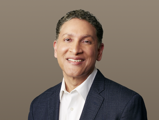
At DLVSC, we have a highly regarded, fellowship-trained Charlotte Mohs surgeon: Dr. Gilly Munavalli. Dr. Munavalli completed his Mohs Micrographic Surgery fellowship at The University of California San Francisco, a program approved by the American College of Mohs Micrographic Surgery and Cutaneous Oncology (ACMS).

Mohs Surgery is performed in our Midtown and Pineville offices on an outpatient basis, under local anesthesia. Below you will find helpful information regarding your Mohs surgery, FAQs and more.

Patient Guide: Mohs Surgery Wound Care

Patient Guide: Before Your Mohs Surgery

Our Mohs Surgeon

Gilly Munavalli, MD, MHS, FACMS
Gilly Munavalli, MD, MHS, FACMSMedical Director | Physician

FAQs

In 1938, University of Wisconsin surgeon Dr. Frederic Mohs (rhymes with nose) first introduced his more sophisticated & effective technique for skin cancer removal, now referred to as Mohs Micrographic Surgery. The technique has been refined since its inception – gaining recognition for both its high cure rates and lowered risk of complications & disfiguring scars. Today, Mohs Micrographic Surgery is widely-accepted across the globe as the gold standard in complex skin cancer removal; cure rates reach as high as 99% for Basal Cell Carcinomas and 95% for Squamous Cell Carcinomas and skin cancers previously treated with another method.

In 1938, University of Wisconsin surgeon Dr. Frederic Mohs (rhymes with nose) first introduced his more sophisticated & effective technique for skin cancer removal, now referred to as Mohs Micrographic Surgery. The technique has been refined since its inception – gaining recognition for both its high cure rates and lowered risk of complications & disfiguring scars. Today, Mohs Micrographic Surgery is widely-accepted across the globe as the gold standard in complex skin cancer removal; cure rates reach as high as 99% for Basal Cell Carcinomas and 95% for Squamous Cell Carcinomas and skin cancers previously treated with another method.

Mohs Micrographic Surgery is indicated for biopsy-proven skin cancers that are large, fast-growing, ill-defined, recurrent or located in areas of the body where recurrences are likely and/or when preservation of tissue is especially important (i.e. nose, eyelids, ears, lips, scalp, hands). Since all skin cancers do not require Mohs surgery, referral for this procedure from a dermatologist or other physician is usually required by health insurance coverage. Mohs surgery may also be used for treating certain aggressive types of skin cancers or in immunosuppressed patients.

It depends. Mohs surgery is a very precise form of skin cancer removal and the full process takes some time. We advise our patients to plan to stay with us for the majority of the day. Please do not schedule unrelated appointments or make other plans on your scheduled surgery day as it will be impossible to predict the exact length of time you will be here.

The Mohs surgery skin cancer removal process is depicted step-by-step in the infographic below. After the skin cancer removal process is complete, Dr. Munavalli will discuss the options for repairing your wound. The most frequently used repairs are direct closure, skin flaps, skin grafts and second intention repairs. These repairs are almost always performed in our Charlotte office immediately following the removal process.

Under rare circumstances, you may be referred to a plastic surgeon for repair.

The Mohs Surgery Process

Step 1: The roots of a skin cancer may extend beyond the visible portion of the tumor. If these roots are not removed, the cancer will recur.

Step 2: The visible portion of the tumor is surgically removed.

Step 3: A layer of skin is removed and divided into sections. The ACMS surgeon then color codes each of these sections with dyes and makes reference marks on the skin to show the source of these sections. A map of the surgical site is then drawn.

Step 4: The undersurface and edges of each section are microscopically examined for evidence of remaining cancer.

Step 5: If cancer cells are found under the microscope, the ACMS surgeon marks their location onto the “map” and returns to the patient to remove another layer of skin – but only from precisely where the cancer cells remain.

Step 6: The removal process stops when there is no longer any evidence of cancer remaining in the surgical site. Because Mohs surgery removes only tissue containing cancer, it ensures that the maximum amount of healthy tissue is kept intact.